Glaucoma is a disease caused by increased intraocular pressure (IOP) resulting either from a malformation or malfunction of the eye's drainage structures. Left untreated, an elevated IOP causes irreversible damage to the optic nerve and retinal fibres resulting in a progressive, permanent loss of vision. However, early detection and treatment can slow, or even halt the progression of the disease.
What causes glaucoma?
The eye constantly produces aqueous, the clear fluid that fills the anterior chamber (the space between the cornea and iris). The aqueous filters out of the anterior chamber through a complex drainage system. The delicate balance between the production and drainage of aqueous determines the eye's intraocular pressure (IOP). Most people's IOPs fall between 8 and 21. However, some eyes can tolerate higher pressures than others. That's why it may be normal for one person to have a higher pressure than another.�
Common types of glaucoma
Open Angle
Open angle (also called chronic open angle or primary open angle) is the most common type of glaucoma. With this type, even though the anterior structures of the eye appear normal, aqueous fluid builds up within the anterior chamber, causing the IOP to become elevated. Left untreated, this may result in permanent damage of the optic nerve and retina. Eye drops are generally prescribed to lower the eye pressure. In some cases, surgery is performed if the IOP cannot be adequately controlled with medical therapy.�
Acute Angle Closure
Only about 10% of the population with glaucoma have this type. Acute angle closure occurs because of an abnormality of the structures in the front of the eye. In most of these cases, the space between the iris and cornea is more narrow than normal, leaving a smaller channel for the aqueous to pass through. If the flow of aqueous becomes completely blocked, the IOP rises sharply, causing a sudden angle closure attack.
While patients with open angle glaucoma don't typically have symptoms, those with angle closure glaucoma may experience severe eye pain accompanied by nausea, blurred vision, haloes around lights, and a red eye. This problem is an emergency and should be treated by an ophthalmologist immediately. If left untreated, severe and permanent loss of vision will occur in a matter of days.
Secondary Glaucoma
This type occurs as a result of another disease or problem within the eye such as: inflammation, trauma, previous surgery, diabetes, tumor, and certain medications. For this type, both the glaucoma and the underlying problem must be treated.
Congenital
This is a rare type of glaucoma that is generally seen in infants. In most cases, surgery is required.
Signs and Symptoms
Glaucoma is an insidious disease because it rarely causes symptoms. Detection and prevention are only possible with routine eye examinations. However, certain types, such as angle closure and congenital, do cause symptoms.
Angle Closure (emergency)
- Sudden decrease of vision
- Extreme eye pain
- Headache
- Nausea and vomiting
- Glare and light sensitivity
Congenital
- Tearing
- Light sensitivity
- Enlargement of the cornea
Detection and Diagnosis
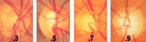 |
| The above photos show progressive optic nerve damage (indicated by the cup to disc ratio) caused by glaucoma.
Notice the pale appearance of the nerve with the 0.9 cup as compared to the nerve with the 0.3 cup. |
Because glaucoma does not cause symptoms in most cases, those who are 40 or older should have an annual examination including a measurement of the intraocular pressure. Those who are glaucoma suspects may need additional testing.
The glaucoma evaluation has several components. In addition to measuring the intraocular pressure, the eye care practitioner will also evaluate the health of the optic nerve (ophthalmoscopy), test the peripheral vision (visual field test), and examine the structures in the front of the eye with a special lens (gonioscopy) before making a diagnosis.
The eye care practitioner evaluates the optic nerve and grades its health by noting the cup to disc ratio. This is simply a comparison of the cup (the depressed area in the center of the nerve) to the entire diameter of the optic nerve. As glaucoma progresses, the area of cupping, or depression, increases. Therefore, a patient with a higher ratio has more damage.
The progression of glaucoma is monitored with a visual field test. This test maps the peripheral vision, allowing the eye care practitioner to determine the extent of vision loss from glaucoma and a measure of the effectiveness of the treatment. The visual field test is periodically repeated to verify that the intraocular pressure is being adequately controlled.
The structures in the front of the eye are normally difficult to see without the help of a special gonioscopy lens. This special mirrored contact lens allows the eye care practitioner to examine the anterior chamber and the eye's drainage system.
Treatment
Most patients with glaucoma require only medication to control the eye pressure. Sometimes, several medications that complement each other are necessary to reduce the pressure adequately.
Surgery is indicated when medical treatment fails to lower the pressure satisfactorily. There are several types of procedures, some involve laser and can be done in the office, others must be performed in the operating theatre. The objective of any glaucoma operation is to allow fluid to drain from the eye more efficiently.
Illustrations by Mark Erickson
With acknowledgement to St. Lukes Eye Hospital.